Figure 1.

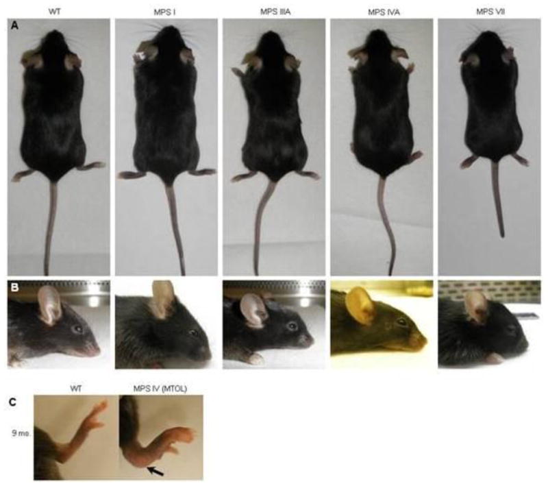
Clinical photographs of MPS I, IIIA, IVA, and VII mice.
A. Full body pictures
B. Side view of head
C. Leg of 9-month-old MPS IVA mouse. Arrow shows abnormal curvature of ankle compared with WT mice

Clinical photographs of MPS I, IIIA, IVA, and VII mice.
A. Full body pictures
B. Side view of head
C. Leg of 9-month-old MPS IVA mouse. Arrow shows abnormal curvature of ankle compared with WT mice