Figure 6.

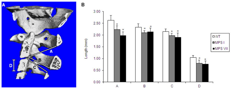
Measurements of dimensions in cervical vertebrae of WT, MPS I, and MPS VII mice.
A. Location of measurements is shown in Fig. 6A.
Line A: From posterior, inferior aspect of the body of cervical vertebrae 2 to the inferior aspect of the arch of cervical vertebrae 1.
Line B: From posterior, inferior aspect of the body of cervical vertebrae 2 to the inferior aspect of the arch of cervical vertebrae 2 (diameter of spinal canal of cervical vertebrae 2).
Line C: From the posterior, superior aspect of the body of cervical vertebrae 3 to the superior aspect of the arch of cervical vertebrae 3 (diameter or spinal canal of cervical vertebrae 3).
Line D: Height of the body of cervical vertebrae 3.
B. Measurements of Lines A–D were made using micro-CT software. Error bars are +/− 1 SD. P-values less than 0.05 compared with WT mice values are represented by an asterisk. Sample sizes are: WT, n=8; MPS I, n=3; MPS VII, n=4 for Line A and B and n=5 for Line C and D.
