
Figure 2. Inhibition of pro-inflammatory cytokine secretion by NOE and SPE in RAW 264.7 macrophages.
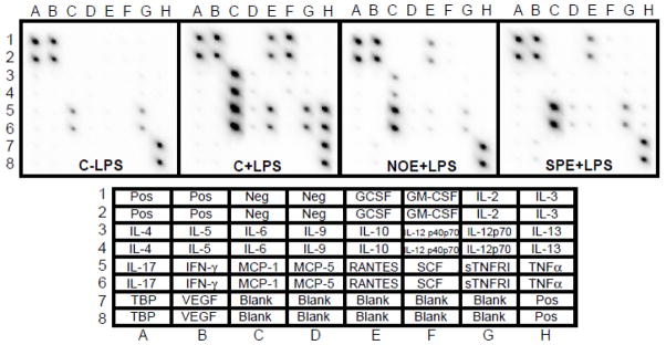
Cells were incubated with 50 μg/mL of NOE or SPE for 12 h and subsequently activated by 100 ng/mL LPS for 18 h. Bottom chart indicates location of cytokines in the array. GCSF, granulocyte colony-stimulating factor; GM-CSF, granulocyte macrophage colony-stimulating factor; IL, interleukins; IFNγ, interferon gamma; MCP-1, monocyte chemoattractant protein 1; RANTES (CCL5), chemokine C-C motive ligand 5; SCF, stem cell factor; sTNFR1, soluble tumor necrosis factor receptor 1; TNFα, tumor necrosis factor alpha; TBP, TNF binding protein; VEGF, vascular endothelial growth factor.