
Figure 6. Alternative mechanisms for anti-inflammatory effects of NOE and SPE in RAW 264.7 macrophages.
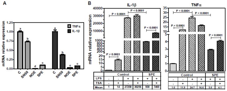
A. Effect of SN50 on pro-inflammatory gene expression. Cells were incubated with NOE or SPE (50 μg/mL) for 12 h. Subsequently, they were incubated with 50 μg/mL of SN50, a NF-κB translocation inhibitor, for 1 h, after which they were activated with 100 ng/mL LPS for 3 h. qRT-PCR analysis was conducted to measure mRNA levels of TNFα and IL-1β. Value are expressed as mean ± SEM, P < 0.05, n = 3. B. RAW 264.7 macrophages were treated with NOE or SPE (100 μg/ml) and 50 nmol/L of TSA for 12 h and then they were activated by LPS (100 ng/mL) for 7 h. qRT-PCR analysis was conducted to measure mRNA levels of TNFα and IL-1β. Value are expressed as mean ± SEM, P < 0.05, n = 3.