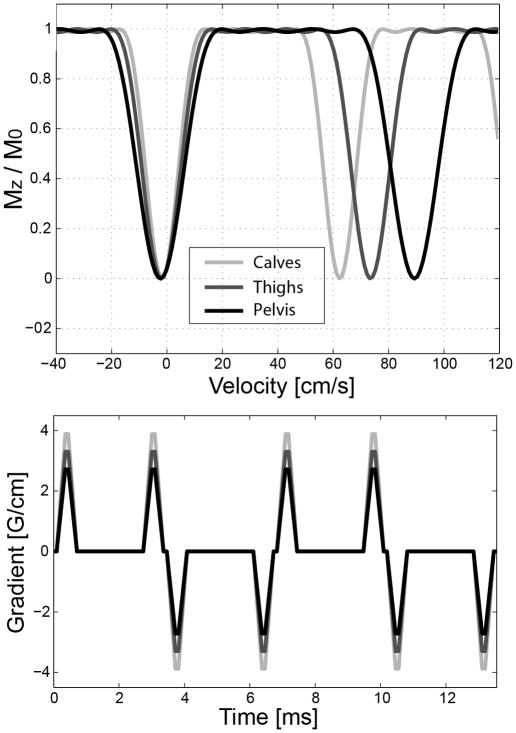
Figure 6.
Simulated Mz profiles of VS saturation pulses (top) and corresponding VS gradient waveforms (bottom) for imaging the calves, thighs and pelvis. The baseline gradient waveform designed for the calves (lightest grey) was scaled down to increase the upper bound of the velocity pass-band for the thighs and pelvis which involve higher arterial flow.

