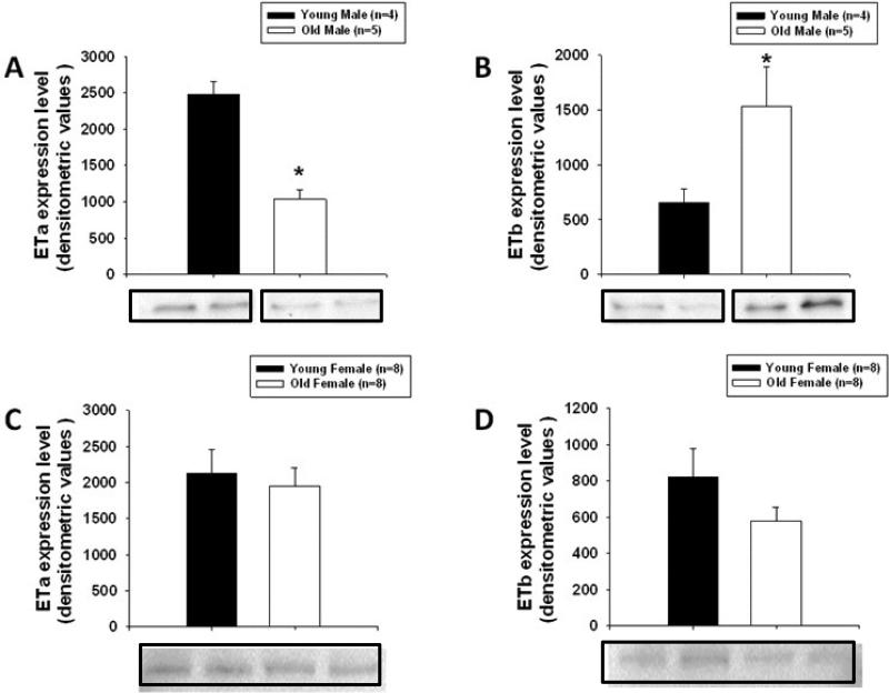
Figure 6.
Advancing age in males caused a decrease in ETa protein expression (A), but an increase in ETb protein expression (B) in coronary arterioles (Young male, n = 4; Old male, n = 5). No age-related differences were found in ETa or ETb protein expression in coronary arterioles from females (C,D) (n = 8 per group). Representative blots of either ETa or ETb receptor protein (~45 kd) are shown below graphs. Equal loading was confirmed by Sypro Ruby staining for total protein. Values are means ± SE. * Indicates significant age-related difference vs. young control, (P ≤ 0.05).

