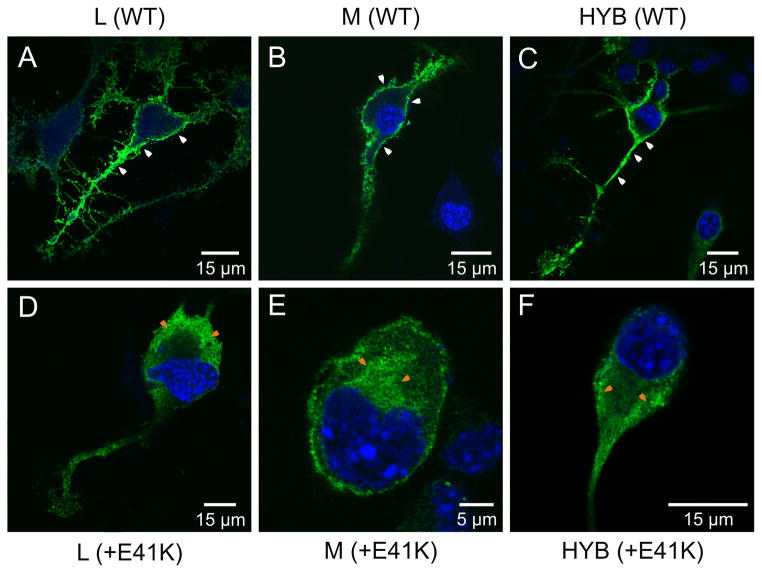
Figure 8.
Confocal microscopy images of transfected Neuro2a cells expressing wild-type and mutant E41K opsins. In all cases, opsin was visualized by the green fluorescence of the Alexa Fluor® 488 dye conjugated to the secondary antibody. (A) L (WT), (B) M (WT), (C) L12M3L4M56 hybrid (HYB), (D) Mutant (E41K) L, (E) Mutant (E41K) M, (F) Mutant (E41K) L1-2M3L4M5-6 hybrid. Note the presence of membrane-associated fluorescence only in cells transfected with the wild-type and hybrid constructs (i.e. without the E41K substitution) (white arrows). Cytoplasmic-associated fluorescence is also indicated (orange arrows).

