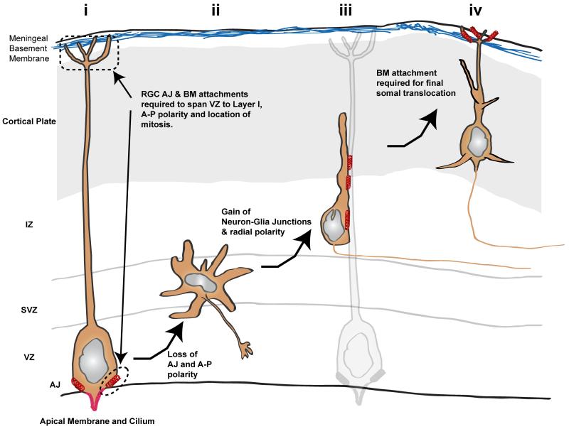
Figure 3.
Schematic illustrating junctional transition, using the developing mouse neocortex as a model. (i) Like epithelial cells, ventricular zone (VZ) RGCs possess strong apical-basal polarity. Their apical AJ contacts and adhesion to basement membrane produced by meningeal cells allow RGCs to span the width of the neocortex. (ii) Both AJs and apical-basal polarity are lost in multipolar neurons in the sub-ventricular zone (SVZ). (iii) Radial migration polarity and neuron-glial cell-cell junctions are re-established in neurons migrating along glial fibers. (iv) Finally, as maturing pyramidal neurons enter the cortical plate, they make Reelin- and N-cadherin–dependent contacts with the marginal zone, facilitating the somal translocation phase of migration. Red springs denote AJ, neuron-glial, or neuron-marginal zone contacts, most of which contain N-cadherin.

