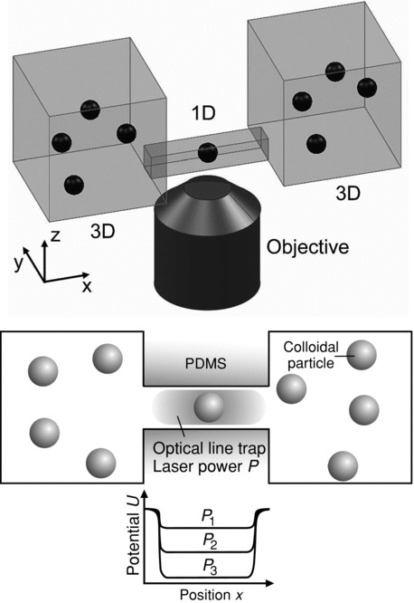
Figure 1.

Illustration of the synthetic membrane channel: Upper panel: two macroscopic reservoirs are connected by a channel allowing for particle transport. Particle diffusion is confined along the channel mimicking quasi-1D diffusion through membrane channels. The diffusion of the particles is quantified by imaging with a 1.4 N.A. 100× objective. Lower panel: simplified top-view of the geometry shown above. Particles diffusing in the channel are subject to an attractive potential from an extended laser line trap generated by holographic optical tweezers. The laser power P can be used to tune the depth of the binding potential in the channel (sketch below the channel) thus mimicking channel-facilitated transport through proteins.
