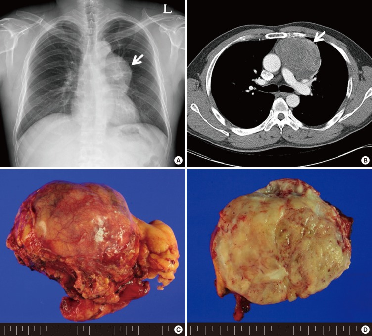
Fig. 1.
Radiologic and gross findings of mediastinal rhabdomyosarcoma arising in mature teratoma. (A) An anterior mediastinal mass in chest X-ray. (B) A 9.5-cm-sized anterior mediastinal mass in Chest Computed tomography. (C) 9.5-cm-sized a well-circumscribed round and firm mass, which is covered as normal thymus and peri-thymic soft tissue. (D) The cut surface of mass show flesh meat like pale tan appearance.

