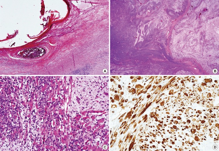
Fig. 2.
Findings of microscopy and immunohistochemistry. (A) In teratoma area, mature squamous epithelium is seen. It is very close to embryonal rhabdomyosarcoma component (H&E, × 40). (B) Low power view of sarcoma area. Intermingled dense compact cellular area and loose myxoid area (H&E, × 12.5). (C) Numerous rhabdoid myoblasts. Epithelioid rhabdomyosarcoma cell showed deeply eosinophilic cytoplasm and small eccentric oval shaped nuclei (H&E, × 400). (D) Tumor cells show immunopositivity for Desmin (× 400).

