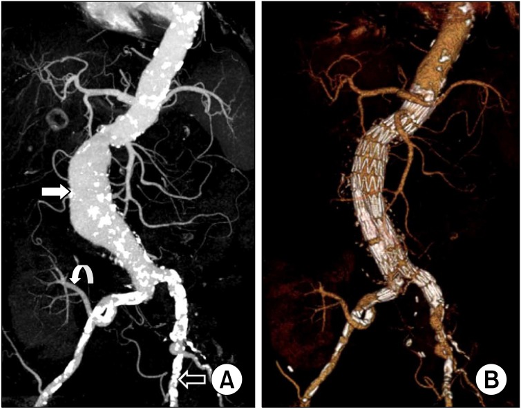
Fig. 2.
Patient II: preoperative and postoperative computed tomography angiography (CTA). (A) The maximal intensity projection view of preoperative CTA showed the abdominal aortic aneurysm (white arrow), the renal artery of functioning transplanted kidney originated right internal iliac artery (curved arrow), and heavily calcified left external iliac artery (open arrow). (B) The volume rendering view of postoperative CTA showed no endoleak and well perfused transplanted kidney.

