
Figure 1. Structure and morphology of hemozoin crystals.
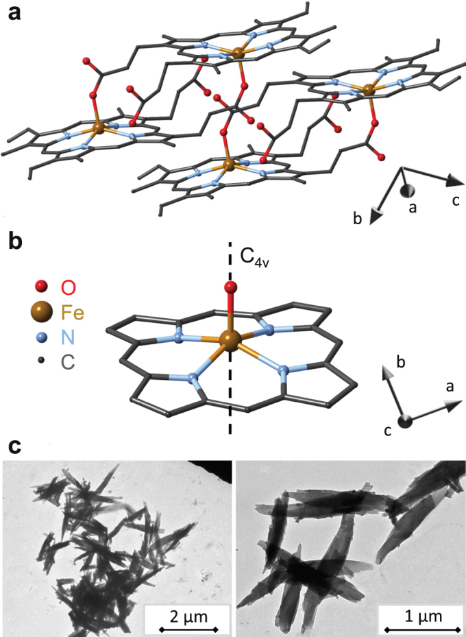
(a) Triclinic structure of hemozoin with two unit cells displayed using structural data from Ref. 29. The main crystallographic axes a, b and c are also indicated. (b) The local symmetry of five-fold coordinated iron in hemozoin nearly preserves a four-fold rotation axis, C4v. The angle spanned by this C4v axis (hard axis of the magnetization) and the crystallographic c-axis (fore-axis of the elongated crystals) is δ ≈ 60°, where the c-axis points out of the plane of the figure. (c) Transmission electron micrographs of typical hemozoin crystallites dried from suspensions.