Figure 6.

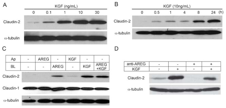
KGF and AREG induce claudin-2 expression predominantly through basolateral signaling. (A) KGF was added to the basolateral compartment of polarized Caco-2 cells in increasing concentrations for 24 hours. Western blot analysis demonstrated that increases in claudin-2 were observed with as low as 0.1 ng/ml KGF. (B) Ten ng/ml KGF was added to the basolateral side of polarized Caco-2 cells and claudin-2 expression was determined over 24 hours. KGF-dependent claudin-2 expression increased with time. (C) AREG (10 ng/ml) and/or KGF (10 ng/ml) were added to either the apical (Ap) or basolateral (BL) medium of polarized Caco-2 cells on Transwell® filters. Claudin-2 levels increased in response to basolateral AREG and/or KGF. Apical KGF had a minor effect on claudin-2 expression and combined AREG and KGF did not enhance claudin-2 expression compared to either growth factor alone. (D) Polarized Caco-2 cells were treated with basolateral KGF, an anti-AREG neutralizing antibody, or both for 24 hours. Neutralization of AREG eliminated basal levels of claudin-2 by western blot analysis. Additionally, AREG neutralization attenuated claudin-2 induction by basolateral KGF.
