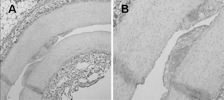
Fig. 9.

The missed soft plaque in two-dimensional images was identified by CD34 immunohistochemistry by its greatly increased brown staining (CD34-positive) in the inner part and base of the plaque compared with adjacent healthy intima. This result indicates a high neovascular density for the plaque, which is consistent with enrichment of the targeted microbubbles in contrast imaging. (a ×40; b ×100). (Color figure online)
