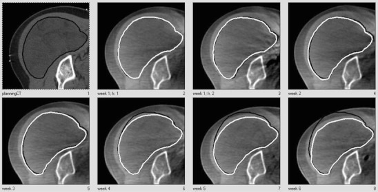
Fig. 1.

Delineations of the GTV of a STS in the buttock, in axial slices. In the upper row, the planning CT and the CBCT scans of the first two fractions of weeks 1 and 2. In the lower row, the CBCT scans acquired in weeks 3 to 6. The delineation of the planning CT scan (black) is projected on each CBCT
