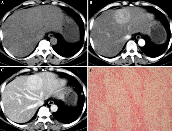
Fig. 3.
56-Year-old woman with epithelioid AML in left lobe of liver (patient 3). A Non-enhanced CT scan shows isoattenuating lesion in segment 4 with fatty liver disease. B Contrast-enhanced CT scan shows obviously enhanced lesion in the arterial phase. C The lesion shows prolonged hyperattenuating in the portal venous phase. D Microscopic examination shows that the tumor is almost exclusively composed of epithelioid cells.

