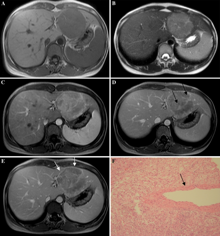
Fig. 4.
32-Year-old woman with epithelioid AML in left lobe of liver (patient 6). A, B: MRI shows a well-defined tumor, hypointense on T1-weighted and hyperintense on T2-weighted images in segment 2/3. C–E: Contrast-enhanced MRI shows inhomogeneous enhanced lesion with punctiform or filiform vessels (arrows) and discrete capsule enhancement (white arrows). F Microscopically, the tumor consists of epithelioid cells with no fat component. There is a thick-walled blood vessel in the center (arrow).

