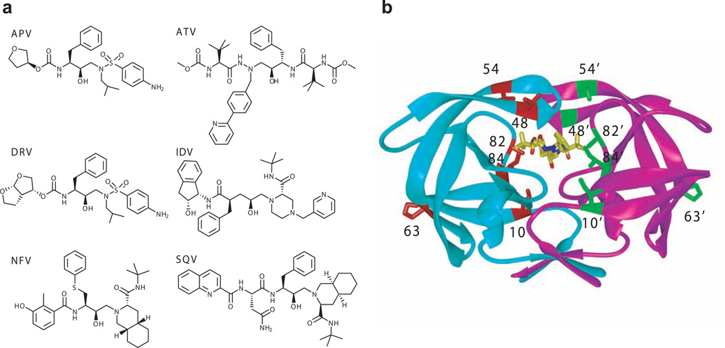
Figure 1.
Structure of inhibitors and HIV-1 protease. (a) Chemical structures of inhibitors. (b) Overview of the mutation sites of Flap+ and Act mutants mapped on an HIV-1 protease dimer. The monomers are distinguished in cyan and magenta, while the inhibitor ATV is shown in yellow stick model. The mutation sites of Flap+ (L10I/G48V/I54V/V82A) and Act (V82T/I84V) along with the site of the natural polymorphism L63P are highlighted in red and green stick models.

