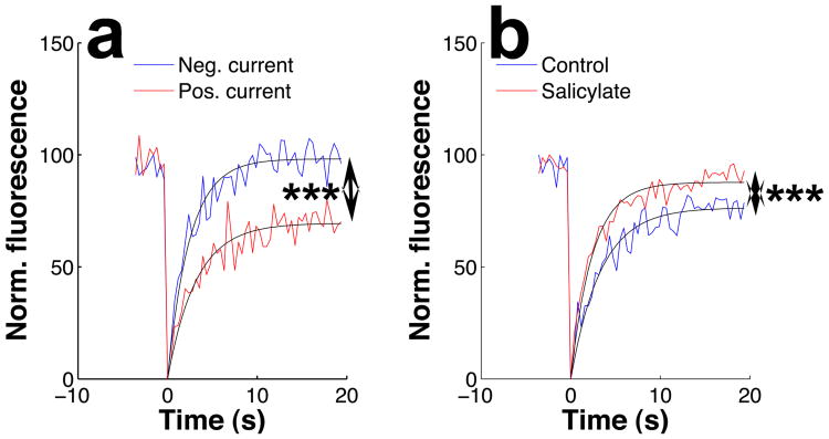
Fig. 5.
Fluorescence recovery after photobleaching in outer hair cell bundles. In each trace, laser light bleached a 1-micron spot on the bundle at time = 0. The fluorescence within the bleached spot is tracked by confocal microscopy before and after bleaching. As dye molecules wander into the bleached area, fluorescence recovers with an exponential time course. a. During negative current stimulation, complete fluorescence recovery is observed, but the extent of recovery is much smaller during positive current. This signifies a decrease in the fraction of dye molecules that are free to move within the membrane. b. Salicylate increases the mobile fraction and the rate of fluorescence recovery. All data in this panel were acquired without current injection. In both panels, smooth lines represent least-squares fits to a single-exponential function. Asterisks signify a p-value less than 0.001 by the Wilcoxon rank sum test.

