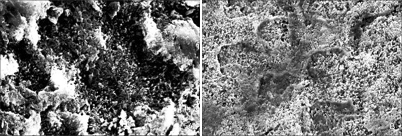
Figure 3.
Scanning electron microscopy images of acid-etched enamel surfaces after 2 weeks of pre-treatment with casein phosphopeptide amorphous calcium phosphate. A, Surface of a tooth etched with 35% phosphoric acid (×5,000). B, Surface of a tooth etched with Transbond Plus Self-Etching Primer (×5,000).

