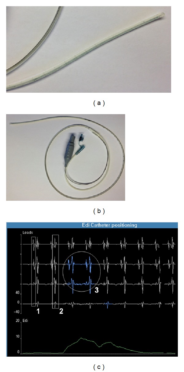
Figure 1.

Specific nasogastric 8F catheter for EAdi monitoring (b), with an enlargement of the distal tip of the catheter equipped with microelectrodes (a). Screenshot of the specific interface for catheter positioning (c) with the three key components of the optimal position: (1) presence of P waves in the proximal lead with disappearance in distal lead; (2) decrease in the QRS amplitude from the upper to the lower leads; and (3) diaphragm electrical activity highlighted mostly in the central leads (in blue).
