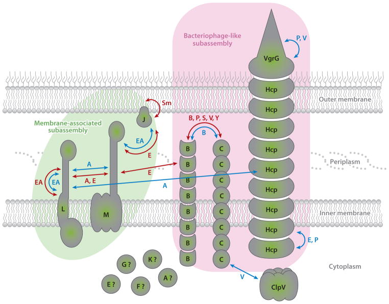
Figure 1.
Protein interaction network between type VI secretion subunits. The localization and topologies of the core components of the T6SS are represented. Arrows indicate interactions detected among the subunits by biochemical/structural (blue) or two-hybrid approaches (red). The letter accompanying the arrow denotes the system where the interaction was detected. The membrane-associated subassembly and the bacteriophage-like subassembly are outlined in green and pink, respectively. The question mark represents subunits for which the localization has not been investigated. Relevant studies are discussed in the text. Abbreviations: T6SS, type VI secretion system; A, Agrobacterium tumefaciens; B, Burkholderia cenocepacia; E, Edwardsiella tarda; EA, enteroaggregative Escherichia coli; P, Pseudomonas aeruginosa; S, Salmonella enterica; Sm, Serratia marcescens; V, Vibrio cholerae; Y, Yersinia pseudotuberculosis Please add references (7, 12, 16a, 31a, 35, 42, 56, 64, 70, 70a, 76, 81, 84, 86 and 116)

