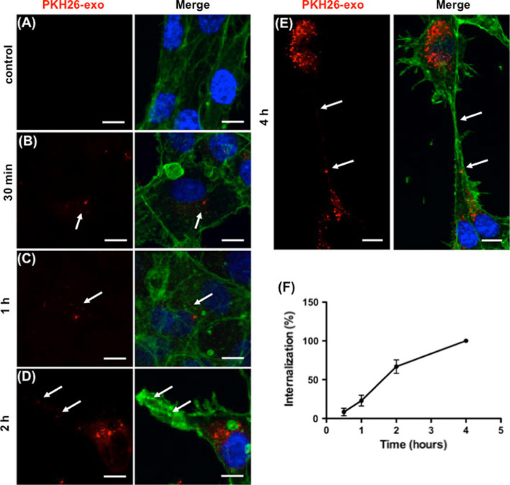
Fig. 3.
Time dependent uptake and localization of exosomes in HUVEC cells during differentiation. a–e Exosomes labeled with PKH26 (red) were added to HUVEC cells at time of plating and incubated as indicated. Effluent from a filtered suspension of PKH26- labeled exosomes was incubated with the HUVECs for 4 h as the control. Cells were fixed and stained for actin (green), and nuclei (DAPI, blue). Arrows indicate exosome localization. Scale bars 10 µm. f Quantitation of HUVEC uptake of PKH26-labeled K562 exosomes. The data represent the mean ± SEM of three independent experiments

