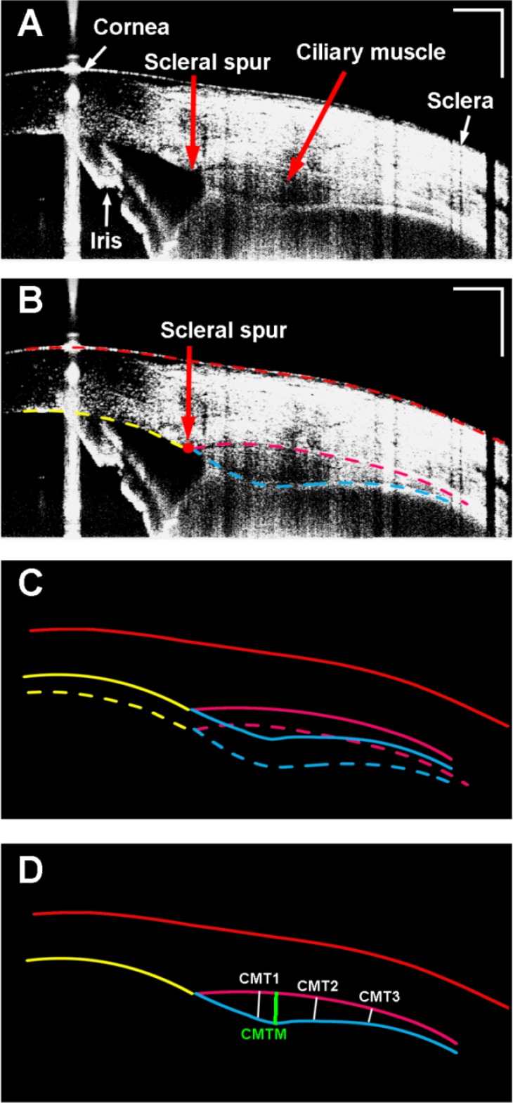
Fig. 4.
Ciliary muscle obtained from a 26-year-old human subject. A: Enhanced image of the ciliary muscle; Note the iris was flipped as the mirror image due to placement of the zero-delay line inside the eye (bottom of the image). B: Semi-automatic segmentation of the boundaries of the cornea, the sclera, and the ciliary muscle. C: Optical correction for image distortion followed Snell’s principle; D: The calculation of the ciliary muscle thickness. CMT1-3: the thickness of the ciliary muscle at 1 mm, 2 mm, and 3 mm posterior to the scleral spur; CMTM: the maximum thickness of the ciliary muscle. Bar = 1 mm.

