
Figure 2. Development of cortical folding in the premature human brain.
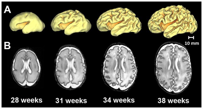
Representative A) 3-dimensional surfaces and B) axial T2-weighted images illustrating regionally-specific cortical folding occurring in the premature brain secondary to sulcation and gyration throughout early development. Provided are images obtained from a single preterm infant from MRI scans performed at 28, 31, 34, and 38 weeks post-mentrual age. Note the marked increase in brain size and folding complexity between each set of images.