
Figure 3. Myelination of the posterior limb of the internal capsule.
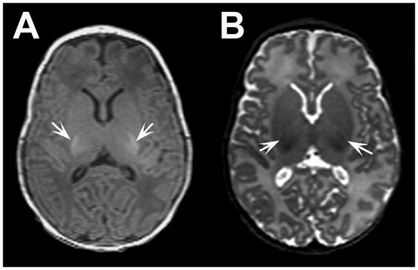
Representative axial A) T1- and B) T2-weighted MR images from a very preterm infant at term equivalent age demonstrating myelination evident in the the posterior limb of the internal capsule bilaterally. Note myelinated white matter appears hyperintense on T1-weighted images and hypointense on T2-weighted images (arrows).