
Figure 4. Classification of periventricular leukomalacia and cerebellar hemorrhage.
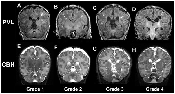
Coronal T1- and T2-weighted MR images demonstrating representative examples of periventricular leukomalacia (PVL, upper panel) and cerebellar hemorrhage (CBH, lower panel) of progressive severity. A) Grade 1 and B) Grade 2 PVL defined by punctate lesions; C) Grade 3 PVL defined by high signal along the wall of lateral ventricles; D) Grade 4 PVL defined by cysts in the periventricular white matter. CBH was also classified into 4 grades including: E) Grade 1 CBH defined by unilateral punctate lesions ≤3 mm in size; F) Grade 2 CBH defined by bilateral punctate lesions; G) Grade 3 CBH defined by a unilateral lesion >3 mm in size; H) Grade 4 CBH defined by extensive lesions bilaterally.