
Figure 5.
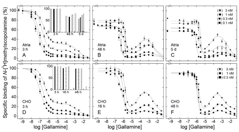
Effect of the concentration of N-[3H]methylscopolamine and the time of incubation on the binding of gallamine to M2 receptor in mammalian membranes. Gallamine and [3H]NMS were mixed simultaneously with membranes from porcine atria (A–C) or CHO cells (D–F), and total binding was measured after incubation of the reaction mixture at 24 °C for different times as follows: porcine atria, 3 h (A), 48 h (B), and 5 d (C); CHO cells, 3 h (D), 16 h (E), and 48 h (F). The concentrations of [3H]NMS and the corresponding levels of occupancy in the absence of gallamine were as follows: 0.1 nM, 51.7% (●); 0.3 nM, 76.4% (■); 1 nM, 91.5% (◆); 3 nM, 97% (★). The lines represent the best fits of Equation 3 (n = 2 or 3) to the data, and the parametric values are listed in Table S7. The asymptotic values of Equation 3 at each time of incubation are compared in the insets to panels A (atrial membranes) and D (CHO membranes) (Y[G]→0, longer bars; Y[G]→∞, shorter bars). The concentration of [3H]NMS was as follows, from left to right: open bars, 0.1 nM; solid bars, 0.3 nM; hatched bars, 1 nM, and checkered bars, 3 nM.