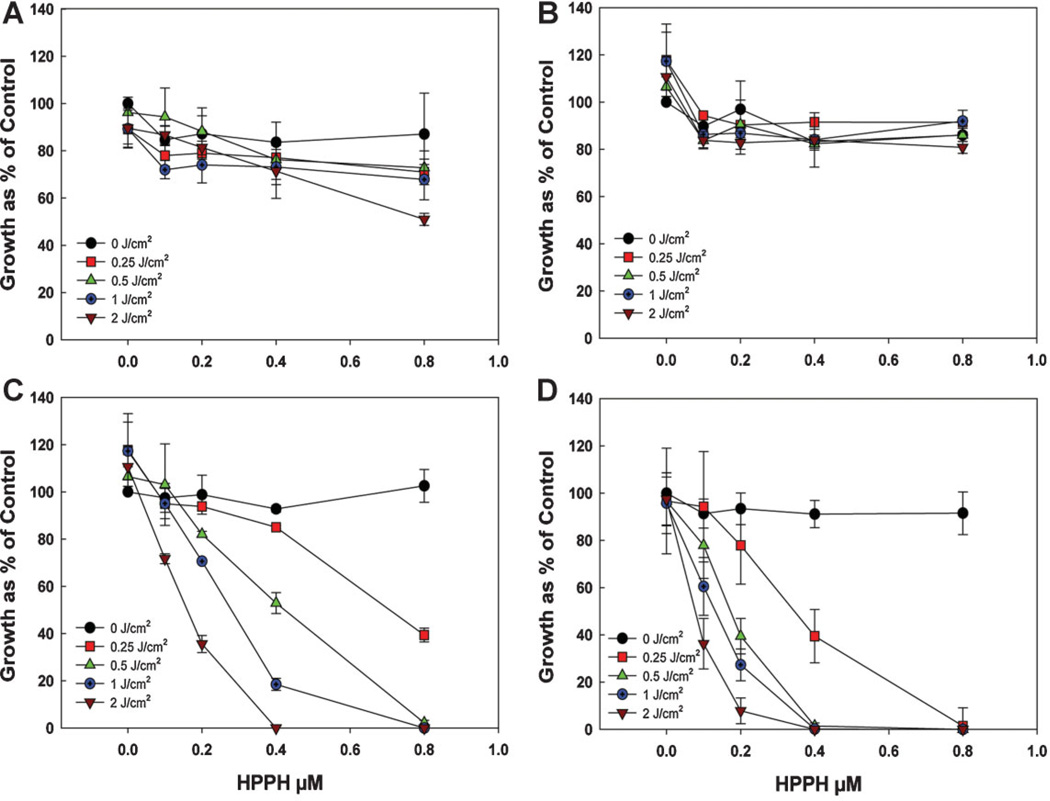
Fig. 5.
Phototoxicity of Colon26 cells after incubation with the different preparations of AFPAA nanoparticles for 4 hours followed by light at a series of doses at a fluence rate of 3.2 mW/cm2. Growth was assayed by the MTT method. (A) Encapsulated HPPH (PAA-E), (B) Conjugated HPPH (PAA-CONJ), (C) Post-loaded HPPH (PAA-PL), (D) Free HPPH. Values are the mean ± standard deviation of 3–4 experiments.

