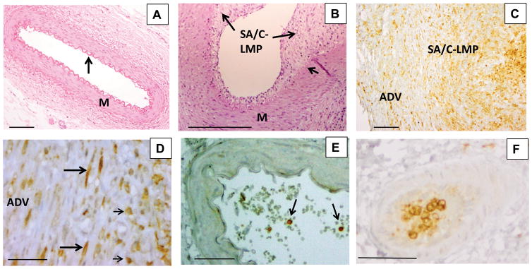
Figure 2.

Histology and immunohistochemistry (IHC) of coronary arteries of KD and childhood control patients. Childhood control coronary artery (A) is free of inflammation and luminal proliferation and has a thin intima covering an undulating elastic lamina (arrow) and a uniform media. Hematoxylin and eosin stain (H&E), 10X objective, scale bar=200 μm. Portion of KD coronary artery (B) with luminal subacute/chronic vasculitis-luminal myofibroblastic proliferation (SA/C-LMP) (long arrows) of varying thickness. The underlying internal elastic lamina (short arrow) is mostly intact, while the media (M) is free of inflammation and somewhat tangentially sectioned. A small portion of visible adventitia in the lower right has subacute/chronic (SA/C) inflammation. H&E 10X objective, scale bar =200μm. IHC for integrin alpha M (ITGAM) (C) reveals positive cells (brown) in the adventitia (ADV), SA/C-LMP, and lumen of a damaged KD coronary artery.10X objective, scale bar =200μm. At higher magnification (D), the positive cells can be seen to be both spindle-shaped (long arrows) and mononuclear inflammatory cells (short arrows). 40X objective, scale bar =50μm. Childhood control coronary artery (E), no ITGAM expression in the arterial wall. A few positive staining circulating mononuclear cells are present in the lumen (arrows). IHC for ITGAM, 20X objective, scale bar =50μm. A section of a small artery located in KD coronary artery periadventitial tissue (F) is almost filled with ITGAM-positive mononuclear cells. IHC for ITGAM, 20X objective, scale bar =50μm.
