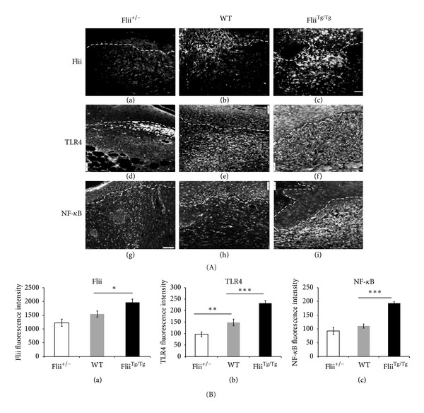
Figure 3.

Concomitant increase in TLR4 and NF-κB staining occurs with increasing levels of Flii. (A) Representative images for Flii (a)–(c), TLR4 (d)–(f), and NF-κB (g)–(i) immunostaining of the three Flii genotypes (Flii+/−, WT, and FliiTg/Tg). (B) Graphical representation of (a) Flii, (b) TLR4, and (c) NF-κB in Flii+/−, WT, and FliiTg/Tg day 7 diabetic wounds. *P ≤ 0.05; **P ≤ 0.01; ***P ≤ 0.001 (n = 6) and scale bar = 100 μm.
