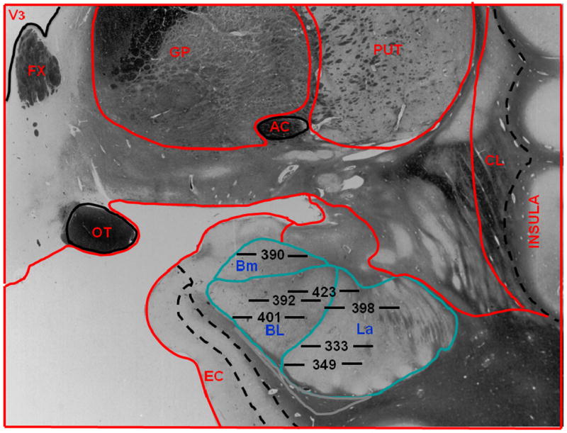
Figure 1.

Location of microdialysis probes in human subjects. Placements of membranes at the tip of each probe are labeled by subject number. Horizontal lines on either side of each subject number are to scale, and their total length indicates the 5.0 mm area sampled by each membrane. The position of each membrane in the amygdala is drawn to scale. The anatomic outline graphic was adapted from the human brain atlas by Mai et al.58. Bm refers to basomedial nucleus of the amygdala; BL, basolateral nucleus of the amygdala, La, lateral nucleus of the amygdala; V3, third ventricle, FX, fornix; GP, globus pallidus; PUT, putamen; CL, claustrum; EC, entorhinal cortex; OT, optic tract; AC, anterior commissure.
