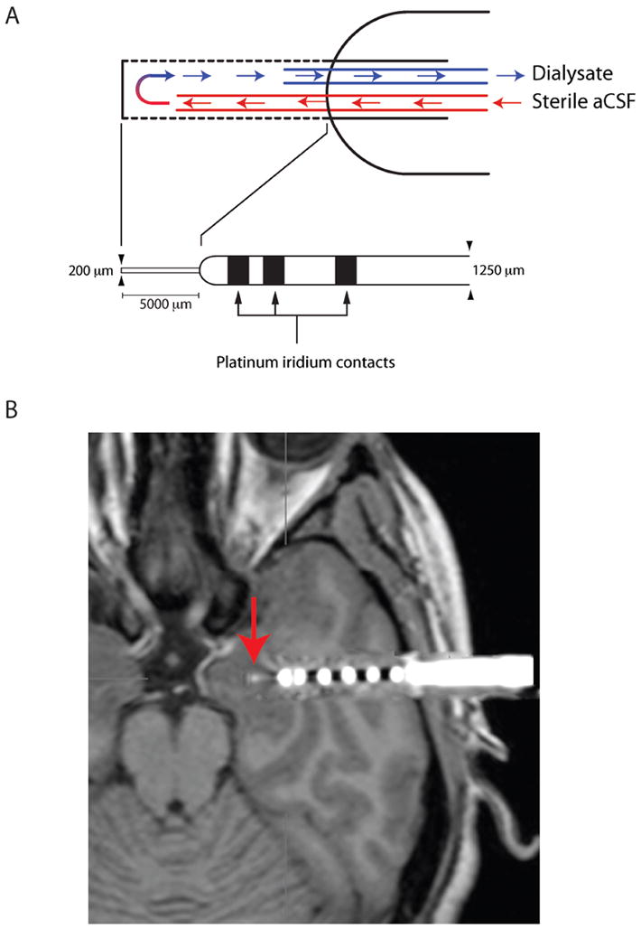
Figure 5.

Design and placement of electrodes. (A) Electrode contained external contacts for localizing seizure focus and internal space for a 200μm diameter microdialysis membrane. The membrane protruded 5 mm from the outer cannula. aCSF flowed in through a 105μm outer diameter silica tube and flowed out of the Cuprophan membrane through a 150μm outer diameter silica tube. Dialysates were collected at 15 min intervals and immediately frozen to -80 degrees. Samples were subsequently analyzed for both Hcrt-1 and MCH using our multiple antigen solid state RIA51. (B) Image of implanted probe in the amygdala (metal contacts produce MRI artifact). CT image of electrode was superimposed on MRI after computer registration (alignment in 3 dimensions) of the two scans.
