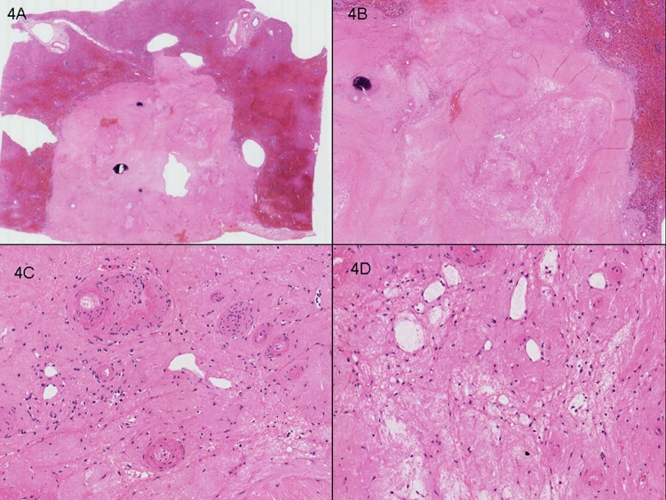
Figure 4.
Case 2. Right hepatectomy with two synchronous tumours. (A) Representative section of lesion 1, 15 mm, showing complete pathological response. (B) No viable tumour cells were seen and the ‘intratumoural’ zone contains marked fibrosis, calcification and paucicellular fibrinous exudate. (C) Ectatic vessels with the larger calibre vessels showing hyalinisation and intimal thickening. (D) Gapping ectatic vessels in association with loose fibrinous exudate and foamy histiocytes.

