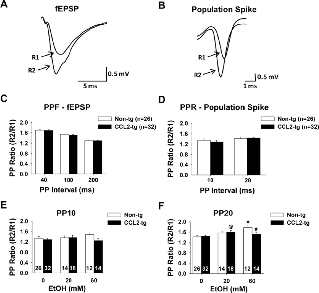
Fig. 4.
Short-term plasticity in CCL2-tg and non-tg hippocampal slices in the absence and presence of acute ethanol. (A-B) Representative traces illustrating paired-pulse facilitation (PPF) of the fEPSP (A) and the PP ratio for the PS (B). Traces R1 and R2 are superimposed responses to paired stimuli separated by 40 ms for the fEPSP and 10 ms for the PS. (C) Summarized results for PPF at 40, 100, and 200 ms paired-pulse intervals. No differences in PPF of the fEPSP slopes were observed for CCL2-tg hippocampal slices compared to non-tg hippocampal slices. (D) Summarized results for the PP ratio at 10 and 20 ms paired-pulse intervals. No differences in the PP ratio for the PS were observed in the CCL2-tg hippocampal slices compared to non-tg hippocampal slices. (E-F) Summarized results for 10 ms (E) and 20 ms (F) paired-pulse intervals for the PS in the presence of 20 mM and 60 mM acute ethanol. Acute ethanol application did not affect the PP ratio of the PS in either CCL2-tg hippocampal slices or non-tg hippocampal slices at the 10 ms interval (E). 20 mM acute ethanol application produced a significant increase in the paired-pulse ratio of the PS in the CCL2-tg hippocampal slices at the 20 ms interval compared with ACSF control CCL2-tg hippocampal slices that were not exposed to ethanol (@, p=0.017). 60 mM acute ethanol application produced a significant increase in the paired-pulse ratio in non-tg hippocampal slices compared with ACSF control non-tg hippocampal slices (*, p=0.004) an effect that was not observed in CCL2-tg hippocampal slices. Thus, there was a significant difference between genotypes in the presence of 60 mM acute ethanol (#, p=0.048). Statistical analysis was determined using the unpaired t-test. The number of slices studied is marked in the corresponding bar.

