Fig. 5.
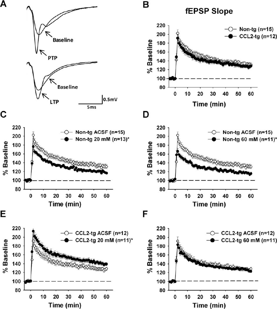
Synaptic plasticity measurements following TBS in hippocampal slices from CCL2-tg and non-tg mice in the absence and presence of acute ethanol. (A) Representative dendritic fEPSP traces illustrating post-tetanic potentiation (PTP) 1–3 minutes following TBS and longterm potentiation (LTP) 50–60 minutes following TBS compared to baseline traces recorded prior to TBS. (B) Synaptic plasticity measurements in hippocampal slices from CCL2-tg and non-tg mice expressed as percent of baseline fEPSP slope. High-frequency stimulation (TBS) was used to elicit synaptic plasticity and occurs at time zero. No differences in synaptic plasticity were observed between CCL2-tg and non-tg hippocampal slices (p=0.968). (C-F) Synaptic plasticity measurements in the presence and absence of acute 20 mM and 60 mM ethanol in non-tg (C and D) and CCL2-tg (E and F) hippocampal slices. A decrease in synaptic plasticity was observed in the presence of 20 mM (C, p < 0.0001) and 60 mM (D, p < 0.0001) ethanol in non-tg hippocampal slices when compared to the ACSF treated non-tg hippocampal slices. An enhancement in synaptic plasticity was observed in the presence of 20 mM ethanol (E) in CCL2-tg hippocampal slices when compared to the ACSF treated CCL2-tg hippocampal slices (p < 0.0001). There was no effect of acute 60 mM ethanol on synaptic plasticity in the CCL2-tg hippocampal slices (F) when compared to the ACSF treated CCL2-tg hippocampal slices (p=0.292). Statistical analysis was determined using repeated measures ANOVA. * Indicates a significant difference between ethanol and ACSF treated hippocampal slices.

