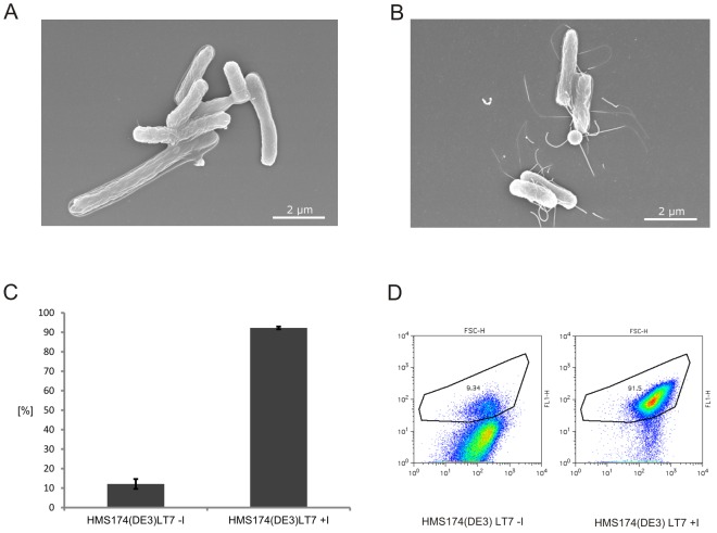
Figure 6. Analysis of the novel host strain HMS174(DE3)LT7.
(A) Scanning Electron Microscopy (SEM) of the HMS174(DE3)LT7 strain without addition of the inducer IPTG. (B) Scanning Electron Microscopy (SEM) of the HMS174(DE3)LT7 strain with addition of the inducer IPTG. Samples were prepared after 90 min cultivation. (C) Cell culture homogeneity. The homogeneity regarding fliC promoter activity of three parallel HMS174(DE3)LT7 strain cultivations containing the plasmid p5′UTR-fliC20-GFPmut-Flag-Linker-StrepII-3′UTR with and without addition of the inducer IPTG were analyzed via Fluorescence Activated Cell Sorting (FACS). After incubation for 2 h at 220 rpm at 37 °C all samples were normalized to OD 600: 1.0 and the percentage of GFP expressing cells indicating flagellar assembly was determined. (D) FACS analysis of GFP expressing cells. A sample of the HMS174(DE3)LT7 strain cultivation containing the plasmid p5′UTR-fliC20-GFPmut-Flag-Linker-StrepII-3′UTR (from Figure 6C) with and without addition of the inducer IPTG.

