
Figure 4. Cardiomyogenic differentiation of Type A and Type B CPC populations.
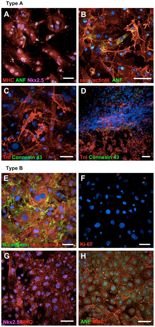
(A and B) Type A CPCs differentiated into cardiomyocytes, when treated with dexamethasone. Organized sarcomeric structures were evident. (C and D) Differentiating cells from Type A CPCs formed multi-layered tissue structure resembling muscle fibers, which expressed TnI and Connexin 43 -gap junctions. (E and F) Type B CPCs differentiated into cardiomyocytes and ceased to proliferate (demonstrated by a lack of Ki-67 staining in majority of cells) when treated with 5 -azacytidine. CD45 expression was confirmed in ∼90% of cells (by FACS) before induction of differentiation. Expression of sarcomeric proteins together with N -cadherin was evident. (G and H) Spontaneous differentiation of Type B CPC population into fast proliferating cells (12th passage). Secreted ANF is seen between the cells. Immunostaining was done three weeks after induction of differentiation. Abbreviations: sarc. actinin = Sarcomeric α-actinin, MHC: Cardiac myosin heavy chain; ANF: Atrial natriuretic factor; TnI: Troponin I. DAPI = blue, bar = 50 µm.