
Figure 1. Detection of cell viability by flow cytometric analysis of BrdU-incorporation.
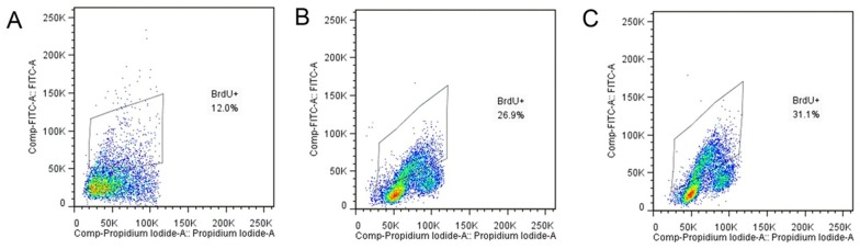
Incorporation of BrdU was measured with a fluorescein isothiocyanate (FITC, green)-conjugated anti-BrdU antibody and propidium iodid (PI, red), shows a typical example of flow cytometric analysis of LoVo cells proliferation after KGF treatment, where a BrdU+ population is clearly visible. Control (A), KGF treated groups with different concentrations including 80 ng/ml (B) and 150 ng/ml (C).