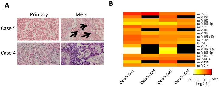
Figure 2. Validation of miRNA expression.
A. In situ hybridization of miR-21. Cancer cells are stained red by Nuclear Red. Higher expression in omental metastases is observed in each case even with both relatively high and low miR-21 expression in the primary tumor. The arrows indicate regions of miR-21 expression co-localizing with Nuclear Red staining. B. Laser Capture Microdissection (LCM) of two cases reveals miRNAs likely expressed in cancer cells. The heat map shows the fold change for the 17 miRNAs identified in the bulk tumor screen. Similar patterns of differential expression are observed for the miRNAs expressed in cancer cells as observed in bulk tumor. Black indicates that the miRNA was not detectable in the LCM isolated cancer cells.

