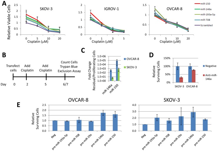
Figure 5. Metastatic miRNAs increase surviving cells.
A. Treatment of cells with pre-miR-150 mimic modestly increases the cisplatin IC50 in SKOV-3 and IGROV-1, but not OVCAR-8 cells. Wst-1 assays were performed 48 hours after cisplatin treatment. Graph shows average of 3 biological replicates. Error bars represent s.e.m. B. Schematic of cisplatin survival assay. Cells are treated twice with high concentrations of cisplatin leading to survival by approximately 1% of the cells. C. miR-150 and miR-146a are up-regulated in surviving cells after 6 days of 50 µM cisplatin in SKOV-3 and 7 days of 30 µM cisplatin in OVCAR-8 cells compared to untreated, proliferating cells. Data are in duplicate and error bars are s.e.m. The fold change for miR-150 is very large because miR-150 was not detectable in proliferating cells. Ct was set to maximum cycle tested, 40, to estimate the fold change. D. Inhibition of miR-146a with 10 nM LNA inhibitor significantly reduces the number of residual cells in SKOV-3 and modestly reduces the surviving cells in OVCAR-8 after 6 days of 50 µM cisplatin in SKOV-3 and 7 days of 30 µM cisplatin in OVCAR-8 cells. The surviving viable cells were determined by trypan blue exclusion assay. Biological triplicate experiments are shown. Error bars are s.e.m. E. Transfection of 50 nM pre-miR-146a and pre-miR-150 increase long term survival after 6 days of 50 µM cisplatin in SKOV-3 and 7 days of 30 µM cisplatin in OVCAR-8 cells.

