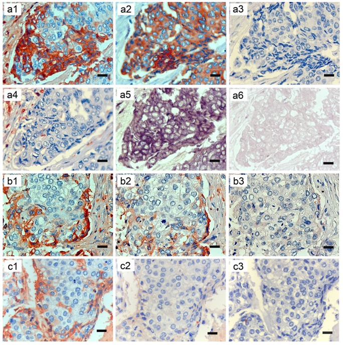
Figure 1. Human breast cancer expresses Igγ protein and IGHG1 mRNA as detected by IHC and ISH.
a1–6, b1–3 and c1–3 are serial sections of breast cancer tissue (IDC) respectively. a1–4 showing IHC with antibodies to to Igγ (IgG heavy chain, a1), CK (an epithelial cell marker, a2), CD20 (a B cell marker, a3) and CD68 (a macrophage marker, a4). a5–6 showing ISH with an IGHG1 mRNA antisense probe (a5), and with a sense probe (a6). b1–3 showing IHC with antibody to Igγ alone, antibody to Igγ preincubated with standard human IgG of 3-fold and 20-fold concentration of the working primary antibody respectively. c1–3 showing IHC staining after incubation with antibody to Igγ, normal rabbit IgG and normal mouse IgG respectively, without positive signals in the Igγ expressing cells in the latter two sections. Original magnifications: ×400. Scale bars: 20 µm.

