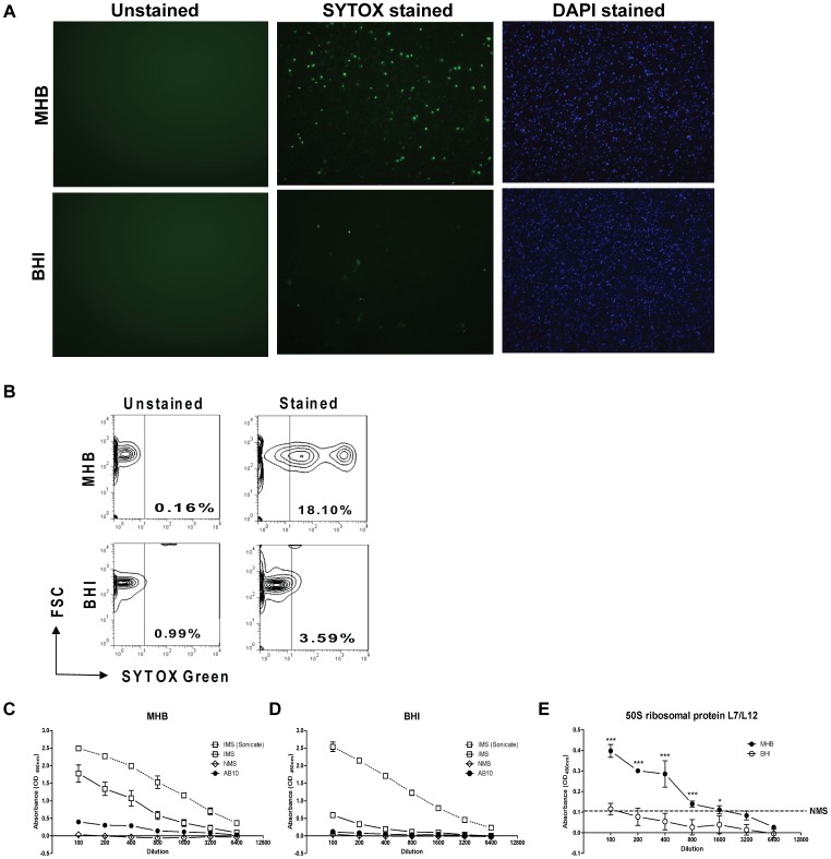
Figure 8. Compared to HAd-Ft, non-HAd Ft are structurally compromised.
(A) The ViaGram™ Red+ bacterial Gram Stain and Viability kit was used to stain HAd-adapted and the non-HAd bacteria. Ft with intact cell membranes stain fluorescent blue with 4′,6-diamidino-2-phenylindole (DAPI), whereas bacteria with damaged membranes stain fluorescent green with SYTOX Green nucleic acid stain, which displaces the DAPI. Ft was visualized using standard fluorescence microscopy. (B) The percentage of structurally compromised bacteria was quantified by flow cytometry. Whole or sonicated (C) non-HAd and (D) HAd-Ft (5×10⁶/well) were coated onto a 96-well microtiter plate and these target antigens were probed with immune sera (IMS) recovered from Ft LVS-infected mice, normal mouse sera (NMS), and a monoclonal antibody (mAb) (AB10) specific for the Ft internal 50S ribosomal protein. Biotinylated anti-mouse IgG mAb was used as a secondary antibody and the plate was developed with TMB substrate (100 µl/well). Reactions were stopped with 2N H2SO4 and absorbance readings were taken at A450 nm. (E) Data for AB10 immune reactivity parsed out from panels (C) and (D) to facilitate comparative analysis. Dotted line indicates limit of detection observed with NMS, which did not differ when antigenic target was MHB- or BHI-grown Ft. *P<0.001, ***P<0.001.

