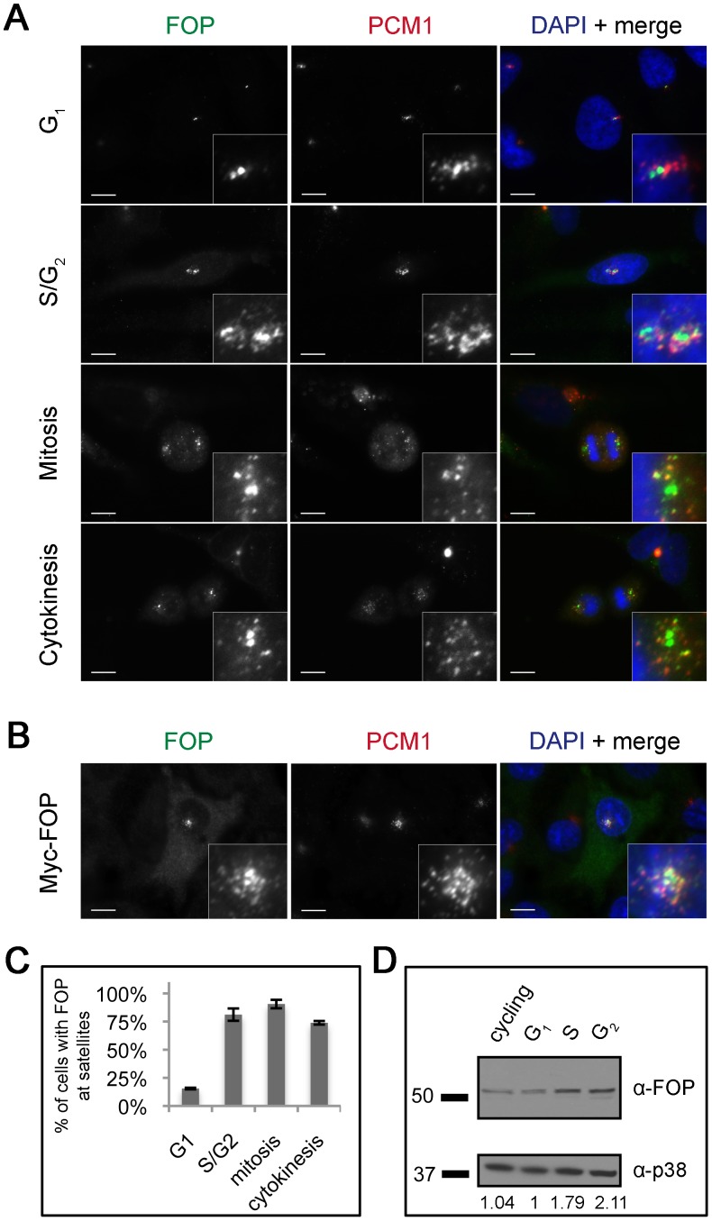
Figure 1. FOP localizes to centrioles and centriolar satellites in a cell cycle-dependent manner.
(A) Asynchronous HeLa cells stained with antibodies against FOP (green) and PCM-1 (red) showing FOP localization at different points in the cell cycle. (B) RPE-1 cells transfected with Myc-FOP and stained with antibodies against Myc (green) and PCM-1 (red). DNA is stained using DAPI (blue). Scale bars: 10 µm; insets: 5× magnification. (C) Quantification of percent of cells with FOP satellite localization during different points in the cell cycle. Bars are mean ± std. dev. from two experiments. Total N = 200, 42, 125, 42, for G1, S/G2, mitosis, and cytokinesis, resp. (D) Western blot analysis of endogenous FOP protein levels in different stages of the cell cycle. Lysates from asynchronous or synchronized HeLa cells were probed with antibodies against FOP and p38 as a loading control. Relative FOP protein levels are calculated as the ratio of FOP/p38 for each lane, normalizing the G1 level to 1.

