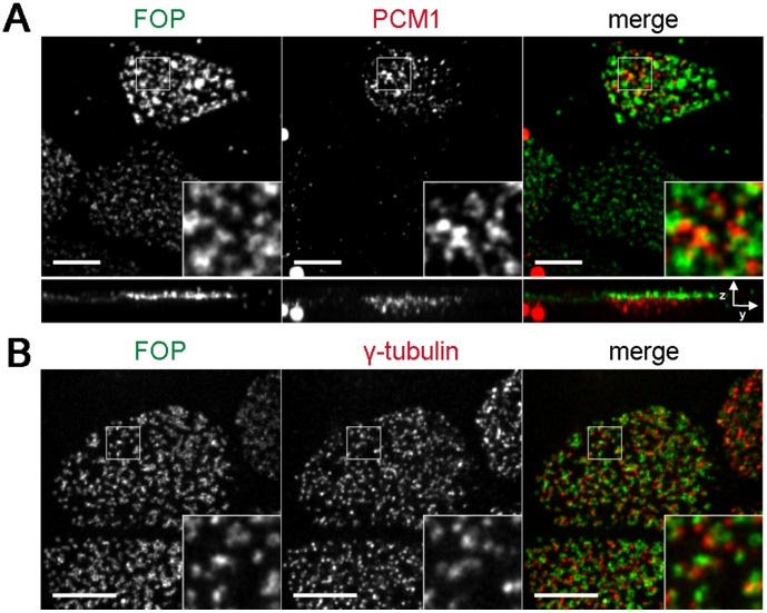
Figure 2. FOP localizes to the basal body layer of multiciliated cells.
Mouse tracheal epithelial cells grown on filters and induced to differentiate by establishment of air-liquid interface. Cells were fixed in paraformaldehyde and stained with antibodies against FOP (green) and PCM-1 (red) to mark satellites (A) or γ-tubulin (red) to mark basal bodies of multiciliated (B). Images shown are maximum projections. Scale bars: 5 and 6 µm resp.; insets: 3× magnification.

