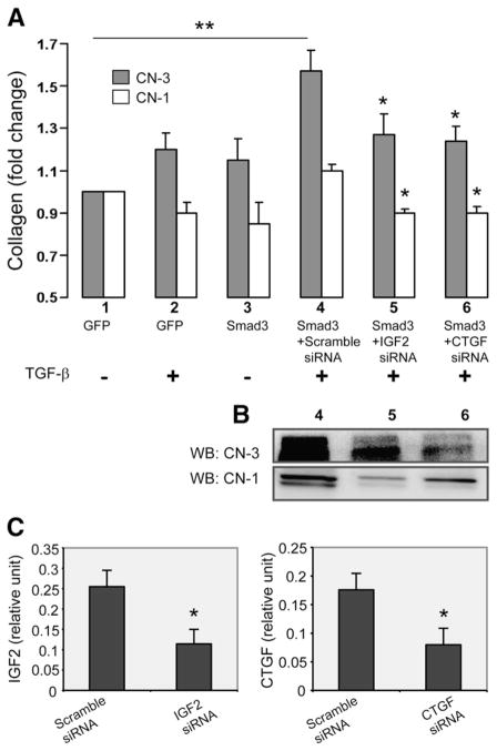
Fig. 5. Effect of CTGF or IGF2 siRNA applied to TGF-β/Smad3-treated SMCs on CN-3 versus CN-1 secretion from adventitial fibroblasts.
A. Measurement of CN-3 and CN-1 by ELISA. Experiments were performed as depicted in Fig. 1, except that respective siRNAs (100 pmol, 6 h) were used to knock down IGF2 or CTGF expression in the Smad3-expressing SMCs. Scramble siRNA (100 pmol) was used as negative control. At 72 h following the addition of the SMC-conditioned media, samples were taken from the fibroblast cultures and analyzed for CN-3 (black) and CN-1 (white) by ELISA. Each bar represents a mean±SEM (at least 3 independent experiments). Different conditions are labeled by numbers (1–6) under the respective bars. **p>0.05, as compared to no TGF-β/Smad3. *p>0.05, as compared to respective maximum CN-3 or CN-1 change (TGF-β/Smad3+scramble siRNA).
B. Western blotting of collagen. In addition to the ELISA measurements in A, CN-3 and CN-1 in the fibroblast media were also detected by immunobotting. Lanes 4, 5, and 6 correspond to the same conditions in A (4, 5, and 6), respectively.
C. Knockdown of IGF2 and CTGF by their respective siRNA, as measured by ELISA. The conditions of scramble siRNA and IGF2 siRNA correspond to those in 4 and 5 in A, respectively; the conditions of scramble siRNA and CTGF siRNA correspond to those in 4 and 6 in A, respectively.

