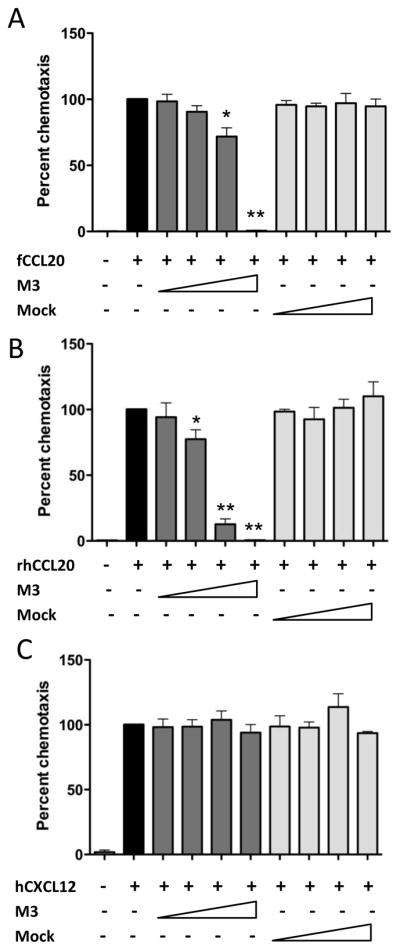
Fig. 6.

M3 protein inhibits CCR6-mediated chemotaxis. (A) Chemotaxis was performed against synthetic fCCL20 (100nM) with or without co-incubation of supernatant from HEK293T cells transiently expressing MHV68 M3 protein (neat, 1:10,1:100 or 1:1000 dilutions) on the bottom side of the membrane separating L1.2.fCCR6 cells from fCCL20. The data are presented as the percent of control chemotaxis observed with fCCL20 alone and are combined from three independent experiments each performed in triplicate. (B) Chemotaxis inhibition was performed as in (A) but with synthetic rhesus macaque CCL20 as the ligand and macaque CCR6-expressing L1.2 cells as the responder cells. (C) Chemotaxis was performed as in (A) but with CXCL12 (1nM) as the ligand and parental L1.2 cells as the responder cells. For all experiments, paired t-test analyses were performed to compare migration in the presence of M3 protein to migration in the absence of M3 protein. P-values of <0.05 (*) and <0.01(**) were observed with the lower dilutions of M3-containing supernatants.
