
Fig. 4.
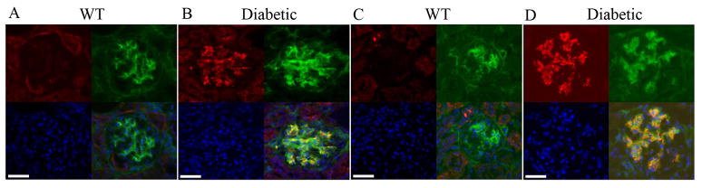
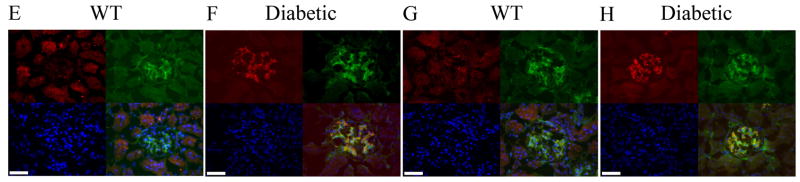
Increased immunoreactivity for PA28 and Cul1/Cul3 proteins in the glomeruli of Ins2Akita/+ mice. No immunoreactivity for PA28-β (red, A) and PA28-γ (red, C) in the 7-month old wild type glomeruli. Ins2Akita/+ mice showed high immunoreactivity for both PA28-β (red, B) and PA28-γ (red, D) which overlapped with PDGF-Rβ labeling of the mesangial cells (green). Immunoreactivity for Cul1 (red, E) and Cul3 (red, G) were determined in 7-month-old wild type (non-diabetic) glomeruli. Ins2Akita/+ mice showed high immunostaining for both Cul1 (red, F) and Cul3 (red, H) which overlaped with PDGF-Rβ labeling of the mesangial cells (green). Nuclei are counterstained with DAPI. Scale bar 25 μm.