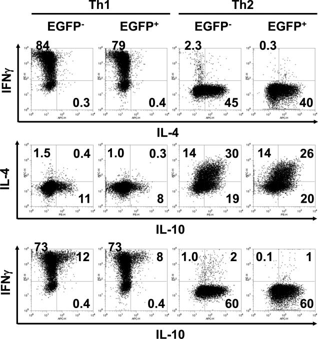
Figure 7. In vitro Th1/Th2 differentiation of CD4 T cells from IL-2cre: Z/EG mouse splenocytes.
Purified naïve CD62Lhigh CD4 T cells were stimulated with plate-bound anti-CD3 plus soluble anti-CD28 antibody under Th1 or Th2-polarizing condition (see materials and methods). After 7 days, cells were re-stimulated PMA and ionomycin for 4 hours and expression of IL-4, IFN- and IL-10 by EGFP+ and EGFP- T cells was analyzed by intracellular cytokine staining. Data are representative of three independent experiments. The numbers indicate the percent of cells within quadrant panels.

